38 eye diagram no labels
Blank ear diagrams and quizzes: The fastest way to learn - Kenhub That's why labeling the ear is an effective way to begin your revision. It helps you to memorize the names and their locations, which in turn will aid you to remember their functions. Below, you can download both the blank ear diagram to make some notes, and then try labeling the ear using the unlabeled ear diagram. Good luck! Wikipedia:Featured picture candidates/Eye-diagram no circles border.svg Eye Diagram Diagram of an Eye On red background to show lost detail. Some parts are white, or white with transparency. Reason Superb quality svg with very high enc value Articles this image appears in Eye Creator Chabacano Nominator Arad Support — Arad 15:05, 8 March 2007 (UTC)
Cow's Eye Dissection - Eye diagram - Exploratorium Learn how to dissect a cow's eye in your classroom. This resource includes: a step-by-step, hints and tips, a cow eye primer, and a glossary of terms. Cow's Eye Dissection - Eye diagram

Eye diagram no labels
The Eye - diagram to label | Teaching Resources The Eye - diagram to label. Subject: Biology. Age range: 14-16. Resource type: Worksheet/Activity. 4.9 13 reviews. canonuk. 4.36842105263158 ... Share through linkedin; Share through facebook; Share through pinterest; File previews. pdf, 2.94 MB. Diagram of eye with key words to use in labelling it. Tes classic free licence. Reviews. 4.9 ... Label Parts of the Human Eye - University of Dayton Parts of the Eye Select the correct label for each part of the eye. The image is taken from above the left eye. Click on the Score button to see how you did. Incorrect answers will be marked in red. File:Eye-diagram no circles border.svg - Wikimedia Commons Description. Eye-diagram no circles border.svg. Afrikaans: 1: Agterste voorportaal 2: Getande rand 3: Siliêre spier 4: Siliêre sonule 5: Schlemm se kanaal 6: Pupil 7: Voorkamer 8: Kornea 9: Iris 10: Lenskorteks 11: Lenskern 12: Siliêre apparaat 13: Konjunktiva 14: Onderste skuinsspier 15: Onderste rektusspier 16: Mediale rektusspier 17 ...
Eye diagram no labels. PDF Parts of the Eye - National Eye Institute | National Eye Institute Eye Diagram Handout Author: National Eye Health Education Program of the National Eye Institute, National Institutes of Health Subject: Handout illustrating parts of the eye Keywords: parts of the eye, eye diagram, vitreous gel, iris, cornea, pupil, lens, optic nerve, macula, retina Created Date: 12/16/2011 12:39:09 PM Eye Anatomy: 16 Parts of the Eye & Their Functions - Vision Center Eye Lens The lens of the eye (or crystalline lens) is the transparent lentil-shaped structure inside your eye. This is the natural lens. It is located behind the iris and to the front of the vitreous humor (vitreous body). The vitreous humor is a clear, colorless, gelatinous mass that fills the gap between the lens and the retina in the eye. BYJUS BYJUS Eye Test: 3 Free Eye Charts to Download and Print at Home Eye doctors can use different eye test charts for different patients and situations. The three most common eye charts are: Snellen eye chart "Tumbling E" eye chart Jaeger eye chart We've included a link to download your very own eye chart after each section below. You can print these charts and test your vision right in your own home.
Eye Diagram Quiz - ProProfs Try this amazing Eye Diagram Quiz quiz which has been attempted 4931 times by avid quiz takers. Also explore over 78 similar quizzes in this category. Take Quizzes. Animal; Nutrition; ... Quiz: Label The Parts Of The Eye. People say that the eyes are the windows to a person's soul. In the class today, we covered parts of the eye, and what ... Human Ear Diagram - Bodytomy The Structure of Human Ear. Helix: It is the prominent outer rim of the external ear. Antihelix: It is the cartilage curve that is situated parallel to the helix. Crus of the Helix: It is the landmark of the outer ear, situated right above the pointy protrusion known as the tragus. Auditory Ossicles: The three small bones in the middle ear ... PDF Eye Anatomy Handout - National Eye Institute of light entering the eye. Lens: The lens is a clear part of the eye behind the iris that helps to focus light, or an image, on the retina. Macula: The macula is the small, sensitive area of the retina that gives central vision. It is located in the center of the retina. Optic nerve: The optic nerve is the largest sensory nerve of the eye. Eye diagram basics: Reading and applying eye diagrams - EDN Eye diagrams provide instant visual data that engineers can use to check the signal integrity of a design and uncover problems early in the design process. Used in conjunction with other measurements such as bit-error rate, an eye diagram can help a designer predict performance and identify possible sources of problems. Also see :
The Eye Diagram: What is it and why is it used? The eye diagram is used primarily to look at digital signals for the purpose of recognizing the effects of distortion and finding its source. To demonstrate using a Tektronix MDO3104 oscilloscope, we connect the AFG output on the back panel to an analog input channel on the front panel and press AFG so a sine wave displays. Then we press Acquire. Label the Eye Worksheet - Teacher-Made Learning Resources - Twinkl Here's a list of the main ones: Iris Sclera Pupil Lacrimal duct Cornea Lens Optic nerve Some of these are visible from the outside, like the iris and the pupil, but others would require a cross-section diagram to even see in the first place. Our Label the Eye worksheet covers all of these and more. Anatomy, medical imaging and e-learning for ... - IMAIOS IMAIOS and selected third parties, use cookies or similar technologies, in particular for audience measurement. Cookies allow us to analyze and store information such as the characteristics of your device as well as certain personal data (e.g., IP addresses, navigation, usage or geolocation data, unique identifiers). Labelling the eye - Science Learning Hub Use your mouse or finger to hover over a box to highlight the part to be named. Drag and drop the text labels onto the boxes next to the eye diagram If you want to redo an answer, click on the box and the answer will go back to the top so you can move it to another box. If you want to check your answers, use the 'Reset incorrect' button.
Anatomy and Physiology I Coursework: Microscope A+P
File:Eye-diagram no circles border 1.svg - Wikimedia Commons Svenska: 1. glaskroppen 2. Ora serrata 3. ciliarmuskeln 4. Zonula ciliaris (kallas ibland zonula-trådar) 5. Schlemms kanal 6. pupill 7. främre ögonkammaren 8. hornhinna (kallas ibland kornea, cornea) 9. iris 10. lins (yttre skikt) 11. lins (kärna) 12. strålkropp 13. bindhinna (kallas ibland konjunktiva) 14.

BBC - GCSE Bitesize: Hurricanes
Labelling the eye — Science Learning Hub Labelling the eye Add to collection The human eye contains structures that allow it to perceive light, movement and colour differences. In this activity, students use online or paper resources to identity and label the main parts of the human eye. By the end of this activity, students should be able to: identify the main parts of the human eye

Eye Label
Label the Eye - The Biology Corner Label the Eye Shannan Muskopf December 30, 2019 This worksheet shows an image of the eye with structures numbered. Students practice labeling the eye or teachers can print this to use as an assessment. There are two versions on the google doc and pdf file, one where the word bank is included and another with no word bank for differentiation.
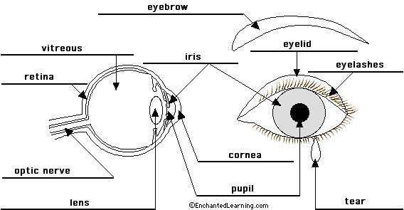
Eye Anatomy Diagram - EnchantedLearning.com
diagram of eye without labels eye diagram human without label anatomy vector labels each medicinebtg Human Skeleton Back No Text No Color Clip Art At Clker.com - Vector skeleton human posterior clip text vector clker svg Relapsing Polychondritis: Causes, Picture, Symptoms And Treatment
Labelled Rib Cage Clip Art at Clker.com - vector clip art online, royalty free & public domain
North Carolina Eye Care & Eye Exams | eyecarecenter North Carolina Eye Care Services. The eye doctors at eyecarecenter are here to help you with all of your eye care needs. Our highly trained eye care professionals focus on maintaining your eye health with comprehensive eye care, preventative care, and treatment. Trust us for routine checkups and treating conditions like glaucoma, cataracts & more.

Eye Diagram: Label Quiz
File:Eye-diagram no circles border.svg - Wikipedia Description. Eye-diagram no circles border.svg. Afrikaans: 1: Agterste voorportaal 2: Getande rand 3: Siliêre spier 4: Siliêre sonule 5: Schlemm se kanaal 6: Pupil 7: Voorkamer 8: Kornea 9: Iris 10: Lenskorteks 11: Lenskern 12: Siliêre apparaat 13: Konjunktiva 14: Onderste skuinsspier 15: Onderste rektusspier 16: Mediale rektusspier 17 ...
Human Body Anatomy Basics No Lines Clip Art at Clker.com - vector clip art online, royalty free ...
Consumer Updates | FDA No FEAR Act; FOIA; HHS.gov; USA.gov; Contact FDA Follow FDA on Facebook Follow FDA on Twitter View FDA videos on YouTube Subscribe to FDA RSS feeds. FDA Homepage. Contact Number 1-888-INFO-FDA (1 ...

The BioLogs: CSEC - The Eye - functions of the various parts
Blank Eye Diagram - Healthiack Best viewed on 1280 x 768 px resolution in any modern browser. Blank eye diagram 1063. Blank eye diagram 1020. Blank eye diagram 1023. Blank eye diagram 1029. Blank eye diagram 1031. Blank eye diagram 1033. Blank eye diagram 1034. Blank eye diagram 1035.
Human Skeleton Back No Text No Color Clip Art at Clker.com - vector clip art online, royalty ...
The Eye and the Ear (Blank) Printable Printable (6th - 12th Grade) Test students' knowledge of the human eye and ear as they color and label these diagrams. Subjects: Science. Human Body and Anatomy. Human Biology.

Human eye - Wikipedia
Generate eye diagram - MATLAB eyediagram - MathWorks eyediagram (x,n,period) sets the labels on the horizontal axis to the range between - period /2 to period /2. eyediagram (x,n,period,offset) specifies the offset for the eye diagram. The function assumes that the ( offset + 1)th value of the signal and every n th value thereafter, occur at times that are integer multiples of period.
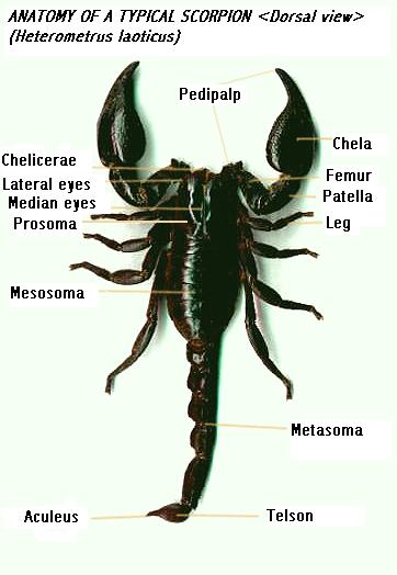
Heterometrus laoticus
Eye Diagram - an overview | ScienceDirect Topics An eye diagram provides a simple and useful tool to visualize intersymbol interference between data bits. Figure 24a shows a perfect eye diagram. A square bit stream (i.e., series of symbol '1's and '0's) is sliced into sub-bit stream with predetermined eye intervals (i.e., several bit periods), and displayed through bit analyzing equipment (e.g., digital channel analyzer), overlapping ...
33 Label The Parts Of The Eye - Labels For You
eye diagram with labels eye diagram with labels Human Skeleton Back No Text No Color Clip Art at Clker.com - vector. 9 Pics about Human Skeleton Back No Text No Color Clip Art at Clker.com - vector : Muscles of the Human Eyeball | ClipArt ETC, Parts of an eye - ESL worksheet by step2eternity and also Human Body Anatomy Basics No Lines Clip Art at Clker.com - vector clip.

quantum art and poetry: Quantum Entanglement explained by time as an emergent property
Vision and Eye Diagram: How We See - AARP Light reflects off the object we're looking at and enters the eye through the cornea, a clear, thin, dome-shaped tissue at the very front of the eye. The cornea has a curvature to it and covers the eye, kind of like a crystal covering the face of a watch. "When rays of light enter the eye, they're sort of parallel to each other," says Rosen.

Correctly Label the Eye Diagram Quiz
File:Eye-diagram no circles border.svg - Wikimedia Commons Description. Eye-diagram no circles border.svg. Afrikaans: 1: Agterste voorportaal 2: Getande rand 3: Siliêre spier 4: Siliêre sonule 5: Schlemm se kanaal 6: Pupil 7: Voorkamer 8: Kornea 9: Iris 10: Lenskorteks 11: Lenskern 12: Siliêre apparaat 13: Konjunktiva 14: Onderste skuinsspier 15: Onderste rektusspier 16: Mediale rektusspier 17 ...
Labeled Eye Diagram - ClipArt Best
Label Parts of the Human Eye - University of Dayton Parts of the Eye Select the correct label for each part of the eye. The image is taken from above the left eye. Click on the Score button to see how you did. Incorrect answers will be marked in red.

Frazer + Biology: 2.86 The Eye
The Eye - diagram to label | Teaching Resources The Eye - diagram to label. Subject: Biology. Age range: 14-16. Resource type: Worksheet/Activity. 4.9 13 reviews. canonuk. 4.36842105263158 ... Share through linkedin; Share through facebook; Share through pinterest; File previews. pdf, 2.94 MB. Diagram of eye with key words to use in labelling it. Tes classic free licence. Reviews. 4.9 ...
Label Eye Diagram
Post a Comment for "38 eye diagram no labels"