44 brain mri with labels
Harvard University Show labels Show list All modalities to: MR-T1 MR-T2 FDG T1/FDG T2/FDG biccn.orgBrain Cell Data Center (BCDC) - Brain Cell Data Center (BCDC) Brain tissues are imaged and spatially registered using MRI, and sampling regions (visual cortex or BA17) are processed for single cell assays. Gene expression and chromatin accessible site mapping permits unbiased clustering of nuclei (visualized using t-SNE) and cell type annotation.
Neuroimaging - Wikipedia Neuroimaging is the use of quantitative (computational) techniques to study the structure and function of the central nervous system, developed as an objective way of scientifically studying the healthy human brain in a non-invasive manner. Increasingly it is also being used for quantitative studies of brain disease and psychiatric illness. Neuroimaging is a highly multidisciplinary …
Brain mri with labels
› AANLIB › casesHarvard University Show labels Show list All modalities to: MR-T1 MR-T2 FDG T1/FDG T2/FDG The Brain | Noba Figure 1. An MRI of the human brain delineating three major structures: the cerebral hemispheres, brain stem, and cerebellum. The brain uses oxygen and glucose, delivered via the blood. The brain is a large consumer of these metabolites, using 20% of the oxygen and calories we consume despite being only 2% of our total weight. However, as long ... Labeled imaging anatomy cases | Radiology Reference Article ... This article lists a series of labeled imaging anatomy cases by body region and modality. Brain CT head: non-contrast axial CT head: non-contrast coronal CT head: non-contrast sagittal CT head: angiogram axial CT head: angiogram coronal CT head: angiogram sagittal CT head: venogram axial CT head: venogram coronal CT head: venogram sagittal
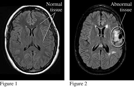
Brain mri with labels. Researchers automate brain MRI image labeling, more than ... - ScienceDaily Researchers have automated brain MRI image labeling, needed to teach machine learning image recognition models, by deriving important labels from radiology reports and accurately assigning them to... nobaproject.com › modules › the-brainThe Brain | Noba Figure 1. An MRI of the human brain delineating three major structures: the cerebral hemispheres, brain stem, and cerebellum. The brain uses oxygen and glucose, delivered via the blood. The brain is a large consumer of these metabolites, using 20% of the oxygen and calories we consume despite being only 2% of our total weight. However, as long ... Brain Cell Data Center (BCDC) - Brain Cell Data Center (BCDC) Retrograde tracers injected into the mouse brain backfill the cell bodies of their input projections and fluoresce, as shown here. ... Surface-based registration is then used to map these labels to models derived ... Brain tissues are imaged and spatially registered using MRI, and sampling regions (visual cortex or BA17) are processed for ... A normative spatiotemporal MRI atlas of the fetal brain for automatic ... Step 2: The segmented neonatal atlases were used to generate initial labels on the spatiotemporal fetal brain MRI atlas at higher GAs (35-37 weeks) through multiatlas segmentation using probabilistic label fusion 65. Step 3: Fetal brain MRI labels were manually defined and propagated in iterations from the higher GAs to the lower GAs.
Brain MRI: What It Is, Purpose, Procedure & Results - Cleveland Clinic A brain (head) MRI scan is a painless test that produces very clear images of the structures inside of your head — mainly, your brain. Healthcare providers use brain MRIs to evaluate, diagnose and monitor several different medical conditions that affect your brain or other structures in your head. Appointments 866.588.2264 Appointments & Locations UCLA Brain Mapping Center - ICBM Template Cortical gyri, subcortical structures and the cerebellum have been delineated from the structural brain template and assigned a unique label. The 3-D set of labels can be imported and registered onto the structural MRI of any individual subject through software like BrainSuite. Brain MRI: How to read MRI brain scan | Kenhub The insular and limbic lobes are the ones of particular interest in the brain MRI. The insular lobe lies just lateral to the extreme capsule of basal ganglia. It is a small portion of the cerebral cortex found deep to the meeting point of the frontal, temporal and parietal lobes. The limbic lobe lies deep to the parietal and frontal lobes. MRI brain (summary) | Radiology Reference Article - Radiopaedia multiplanar assessment of the brain exceptionally detailed images of the brain different sequences allow assessment of different pathology no ionizing radiation (especially important in children) limitations much longer investigation (20-40 minutes) less available (longer waiting list) patients may be claustrophobic
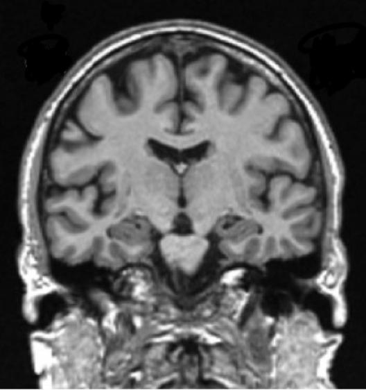
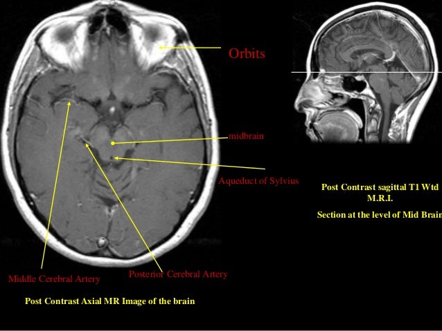
brain anatomy | MRI coronal brain anatomy | free MRI cross sectional ... ELBOW AXIAL. WRIST AXIAL. WRIST CORONAL. KNEE CORONAL. KNEE SAGITTAL. ARTERIES UPPER LEG. ARTERIES LOWER LEG. This MRI brain coronal cross sectional anatomy tool is absolutely free to use. Use the mouse scroll wheel to move the images up and down alternatively use the tiny arrows (>>) on both side of the image to move the images. Brain: Atlas of human anatomy with MRI - e-Anatomy - IMAIOS Sep 13, 2021 · MRI Atlas of the Brain. This page presents a comprehensive series of labeled axial, sagittal and coronal images from a normal human brain magnetic resonance imaging exam. This MRI brain cross-sectional anatomy tool serves as a reference atlas to guide radiologists and researchers in the accurate identification of the brain structures. Labeled MRI Brain Scans - Neuromorphometrics We can also label scans that you provide and we are very interested in labeling white matter anatomy as seen in diffusion-weighted MRI scans. If you want an aggregate version of our data, we can provide it as a probabilistic atlas. The cost to label a single scan is $2449 (USD). MRI head axial T2 - labeling questions - Radiopaedia The labeled structures are (excluding the correct side): cervical spinal cord posterior arch of C1 odontoid process (peg or dens) of C2 parotid gland intradural segment (V4) of dominant vertebral artery cisterna magna intradural segment (V4) of non-dominant vertebral artery cerebellar tonsil occipital condyle medulla oblongata
Clinical MRI Brain Scans Will Enrich the Adult Changes in Thought Study Data Resource - Memory ...
Diffusion MRI - Wikipedia Diffusion-weighted magnetic resonance imaging (DWI or DW-MRI) is the use of specific MRI sequences as well as software that generates images from the resulting data that uses the diffusion of water molecules to generate contrast in MR images. It allows the mapping of the diffusion process of molecules, mainly water, in biological tissues, in vivo and non-invasively.

The Playful Spirit: Functional MRI... cool brain mapping!
Brain lobes - annotated MRI | Radiology Case | Radiopaedia.org mri Axial Coronal Sagittal 3D reconstruction MRI Axial Brain MRI with annotations of major structures. 13 articles feature images from this case 101 public playlists include this case Promoted articles (advertising) Headline: HIV-associated wasting prevalence in the era of modern antiretroviral therapy Javeeda Siddiqui et al., AIDS, 2022
MRI Brain: Purpose, Precautions, Risks & Cost (50% Off Offer)
Reliability-based robust multi-atlas label fusion for brain MRI ... Label fusion is one of the key steps in multi-atlas based segmentation of structural magnetic resonance (MR) images. Although a number of label fusion methods have been developed in literature, most of those existing methods fail to address two important problems, i.e., (1) compared with boundary voxels, inner voxels usually have higher probability (or reliability) to be correctly segmented ...

Dr Balaji Anvekar FRCR: Semi lobar holoprosencephaly MRI
en.wikipedia.org › wiki › Cerebral_cortexCerebral cortex - Wikipedia In the human brain it is between two and three or four millimetres thick, and makes up 40 per cent of the brain's mass. [3] 90 per cent of the cerebral cortex is the six-layered neocortex with the other 10 per cent made up of allocortex . [3]

Figure/Graphic 2a-b: Brain MRI
CPT Code for MRI Brain, Breast, Lumbar Spine and Shoulder Find below the latest Radiology CPT codes for for MRI of Brain, Breast, Lumbar Spine and Shoulder: CPT Codes for MRI Lumbar spine In human Lumbar spine is represented by the 5 vertebrae in between the ribcage and the pelvis forming the largest segment of the vertebral column. Depending on the condition that one is treated on these parts of the ...
MRI SECTIONAL ANATOMY OF BRAIN
Atlas of BRAIN MRI - W-Radiology The most common MRI sequences used include T1-weighted (T1w) and T2-weighted (T2w) scans (5). T1w sequences display those structures mainly made with fat. Thus, they reveal gray matter as gray, white matter as white, bones as black, and cerebrospinal fluid as black. Meanwhile, T2w sequences highlight structures containing more water.
![Untitled Document [www.swjpcc.com]](http://www.swjpcc.com/storage/manuscripts/volume-11/issue-2-august-2015/100-15/100-15 Figure 3.gif)
Untitled Document [www.swjpcc.com]
A multi-atlas label fusion tool for neonatal brain MRI parcellation and ... Introduction. Structure-by-structure analysis, in which the brain is parcellated based on structural units that follow standard ontology in brain anatomy, is widely used to investigate disease-related changes seen on brain MRI scans. 1-4 Numerous tools for brain parcellation methods have been proposed in the past and their accuracy has continuously improved, especially in the past decade ...
Axial Images, Level 4: CT Scan Overview: Chest -- Health Center, University of Connecticut
› en › e-AnatomyBrain: Atlas of human anatomy with MRI - e-Anatomy - IMAIOS Sep 13, 2021 · MRI Atlas of the Brain. This page presents a comprehensive series of labeled axial, sagittal and coronal images from a normal human brain magnetic resonance imaging exam. This MRI brain cross-sectional anatomy tool serves as a reference atlas to guide radiologists and researchers in the accurate identification of the brain structures.

Dr Balaji Anvekar FRCR: Huntington's Disease MRI
MRI anatomy | free MRI axial brain anatomy - Mrimaster.com ELBOW AXIAL. WRIST AXIAL. WRIST CORONAL. KNEE CORONAL. KNEE SAGITTAL. ARTERIES UPPER LEG. ARTERIES LOWER LEG. This MRI brain cross sectional anatomy tool is absolutely free to use. Use the mouse scroll wheel to move the images up and down alternatively use the tiny arrows (>>) on both side of the image to move the images.

Mouse brain seen in sharpest detail ever « Kurzweil
Weight Loss & Diet Plans - Find healthy diet plans and helpful ... - WebMD From healthy diet plans to helpful weight loss tools, here you'll find WebMD's latest diet news and information.

Bio Blog: Technology in Medicine
Cross-sectional anatomy of the brain - e-Anatomy - IMAIOS Anatomical structures and specific areas are visible as interactive labeled images. Cross sectional anatomy: MRI of the brain An MRI was performed on a healthy subject, with several acquisitions with different weightings: spin-echo T1, T2 and FLAIR, T2 gradient-echo, diffusion, and T1 after gadolinium injection.

mri cervical spine sagittal anatomy
Deep learning from MRI-derived labels enables automatic brain tissue ... Deep learning from MRI-derived labels enables automatic brain tissue classification on human brain CT Neuroimage. 2021 Dec 1;244:118606. doi: 10.1016/j ... Our proposed model predicted brain tissue classes accurately from unseen CT images (Dice coefficients of 0.79, 0.82, 0.75, 0.93 and 0.98 for GM, WM, CSF, brain volume and ICV, respectively). ...

Figure/Graphic 1a-b: Brain MRI
› Lower-Limb › Lower-extremity-MRILower extremity: MRI anatomical atlas - e-Anatomy - IMAIOS Feb 07, 2022 · This cross-sectional human anatomy atlas of the lower limb is an interactive tool based on MRI axial images of the human leg. Anatomical structures of the lower limb (hip, thigh, knee, leg, ankle and foot) and specific regions (compartment of the lower limb) are visible on dynamic labeled images.

Nasopharynx Cancer Anatomy and Images
en.wikipedia.org › wiki › NeuroimagingNeuroimaging - Wikipedia Neuroimaging is the use of quantitative (computational) techniques to study the structure and function of the central nervous system, developed as an objective way of scientifically studying the healthy human brain in a non-invasive manner.
Warfarin withdrawal in atrial fibrillation patients awaiting surgery dramatically ups stroke ...
MRI Lables : Merry X-Ray - MXR Imaging MRI Labels. 6 Items. Sort By. Green MRI-Safe Labels. Red MRI Caution Labels 12/PK. Green Safe AND Red Caution Labels 50 COMBO PACK. Green Safe AND Red Caution Labels 24 COMBO PACK. Red MRI Caution Labels 25/PK.

Brain Differences in College Aged Occasional Drug Users - Neuroscience News
Cerebral cortex - Wikipedia The cerebral cortex is the outer covering of the surfaces of the cerebral hemispheres and is folded into peaks called gyri, and grooves called sulci.In the human brain it is between two and three or four millimetres thick, and makes up 40 per cent of the brain's mass. 90 per cent of the cerebral cortex is the six-layered neocortex with the other 10 per cent made up of allocortex.

Neuroanatomy - encyclopedia article - Citizendium
Lower extremity: MRI anatomical atlas - e-Anatomy - IMAIOS Feb 07, 2022 · This cross-sectional human anatomy atlas of the lower limb is an interactive tool based on MRI axial images of the human leg. Anatomical structures of the lower limb (hip, thigh, knee, leg, ankle and foot) and specific regions (compartment of the lower limb) are visible on dynamic labeled images.
Post a Comment for "44 brain mri with labels"