40 brain pictures and labels
Illustration Picture of Brain Anatomy - Brain - eMedicineHealth Medical Illustrations Picture of Brain The brain is the complex organ responsible for processing sensory information (sound, touch, taste, sight, and smell). The brain controls voluntary and involuntary movements. Signals from the brain tell muscles to contract. Input from the brain controls the function of other organs in the body. e-Anatomy: radiologic anatomy atlas of the human body - e-Anatomy - IMAIOS e-Anatomy is an award-winning interactive atlas of human anatomy. It is the most complete reference of human anatomy available on web, iPad, iPhone and android devices. Explore over 6,700 anatomic structures and more than 870,000 translated medical labels. Images in: CT, MRI, Radiographs, Anatomic diagrams and nuclear images. Available in 12 ...
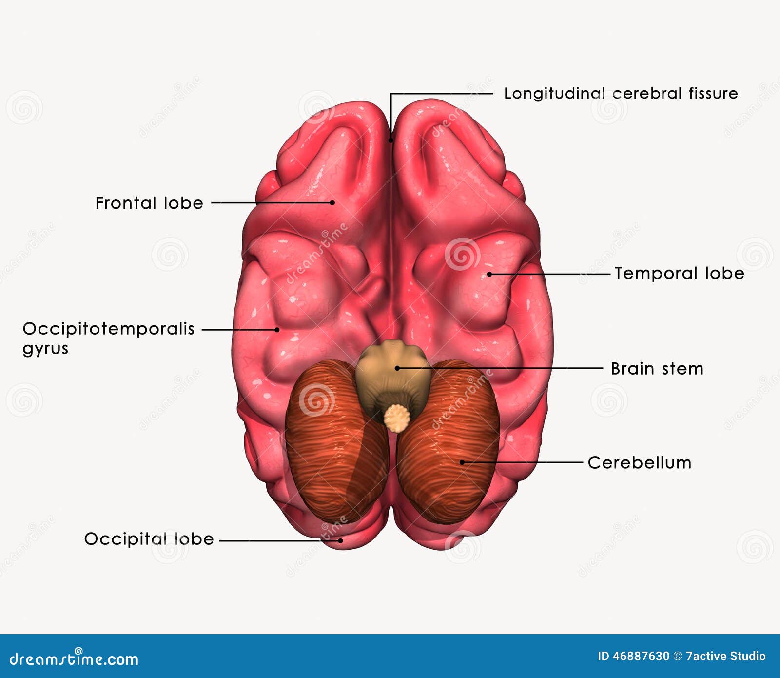
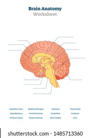
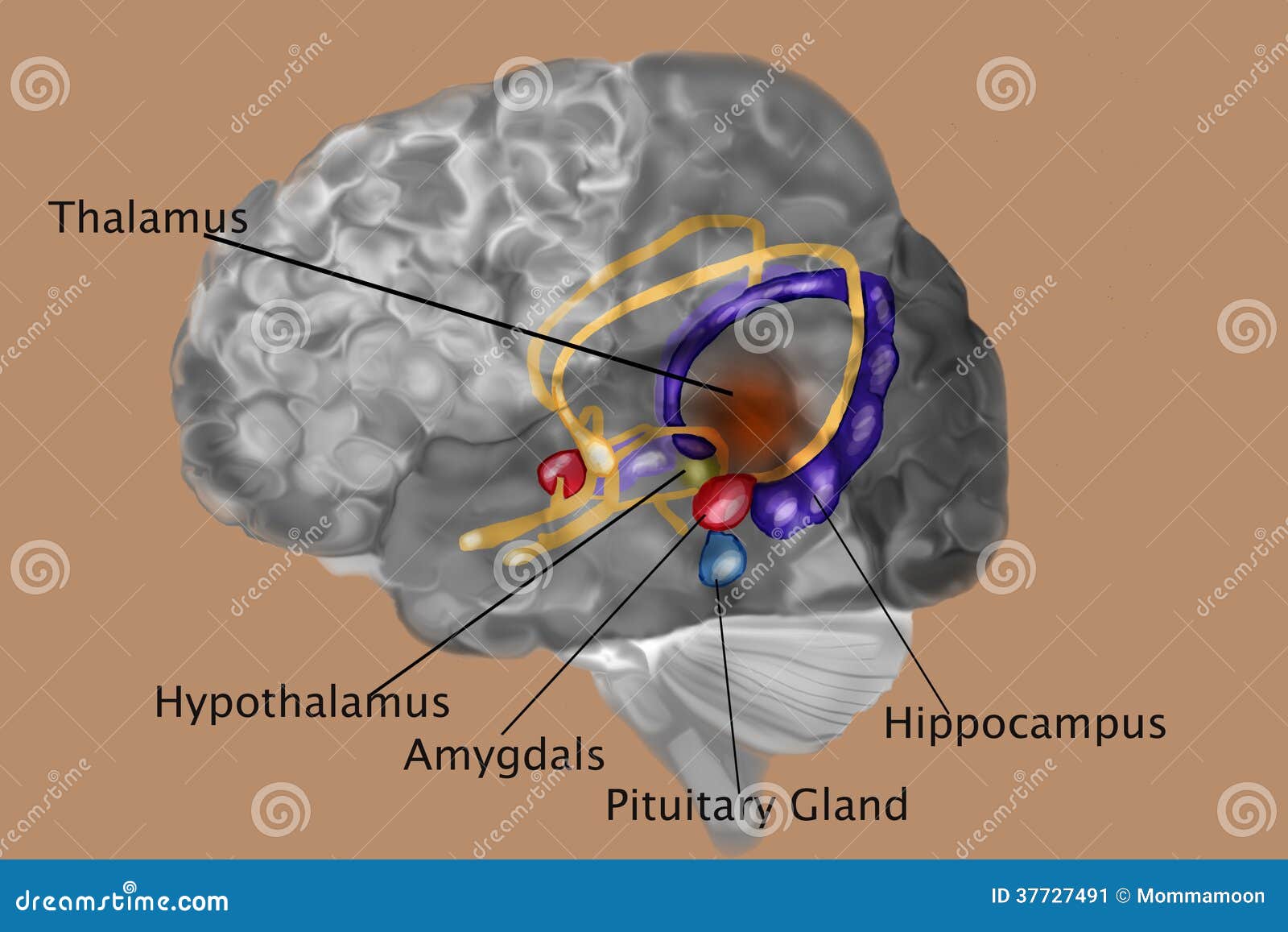
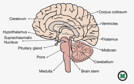
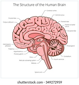
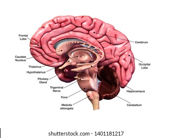
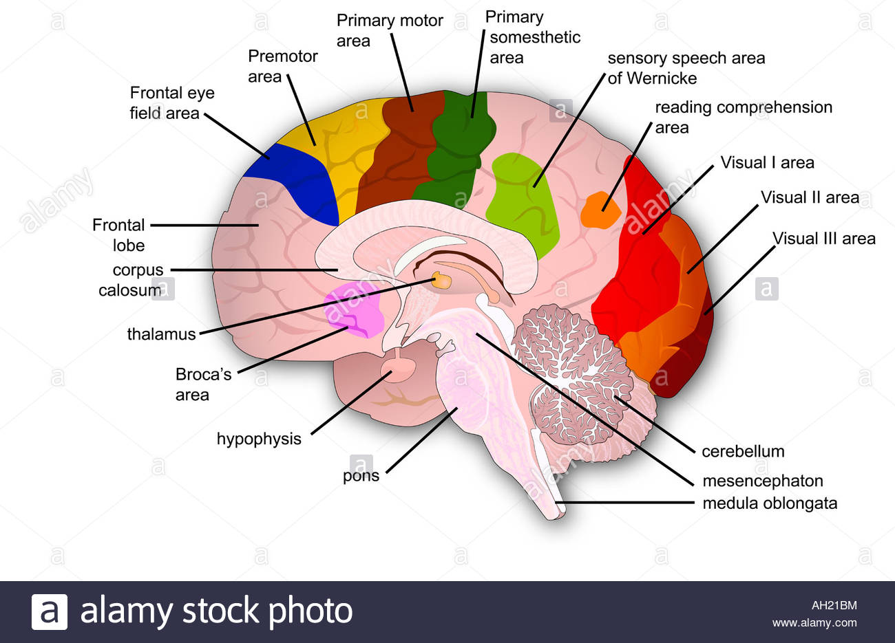
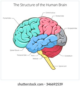
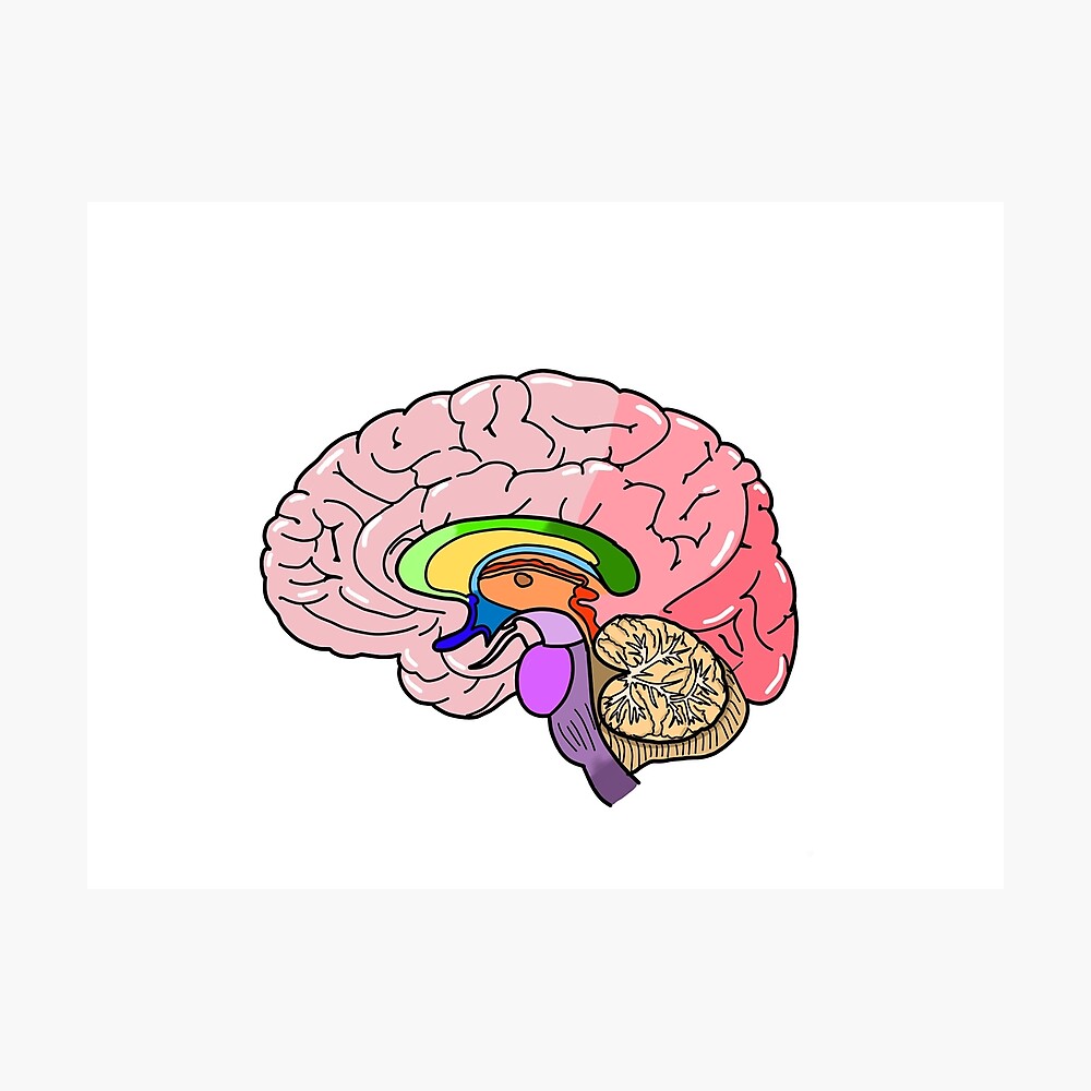
Labeled Diagrams of the Human Brain You'll Want to Copy Now Labeled Diagrams of the Human Brain Central Core The central core consists of the thalamus, pons, cerebellum, reticular formation and medulla. These five regions are the central areas that regulate breathing, pulse, arousal, balance, sleep and early stages of processing sensory information.
Brain pictures and labels
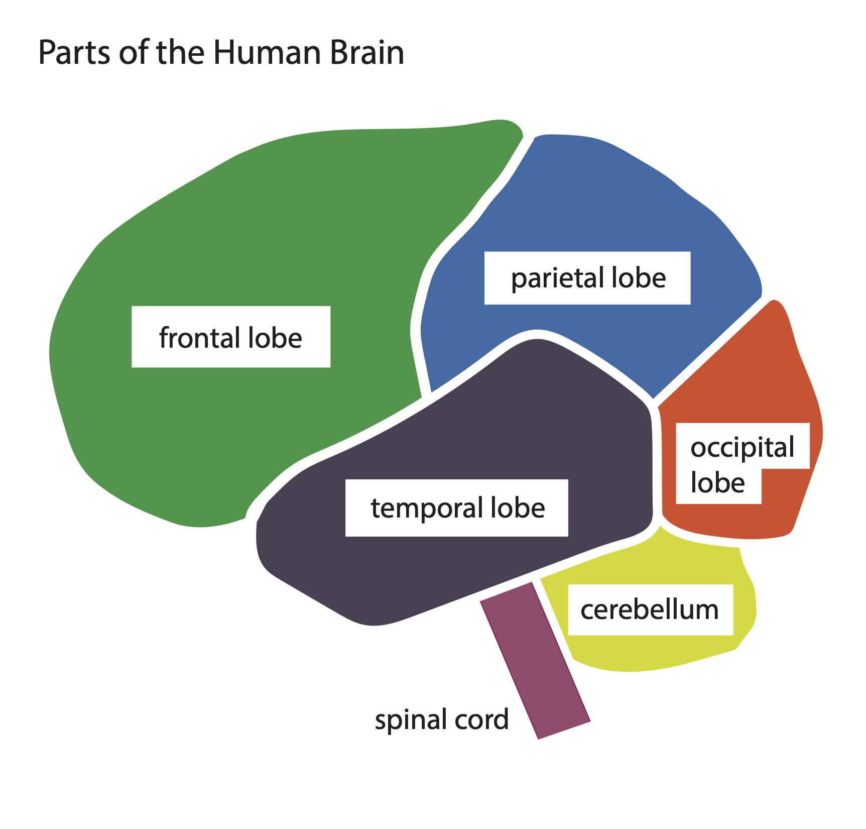
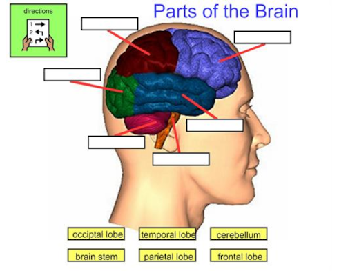
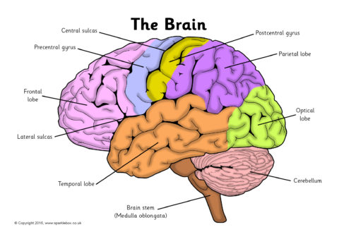
101 Labeled Brain Images and a Consistent Human Cortical Labeling ... We selected 101 T 1-weighted brain MR images that are: (1) publicly accessible with a non-restrictive license, (2) from healthy participants, (3) of high quality to ensure good surface reconstruction, and (4) part of a multi-modal acquisition ( T 2*-weighted, diffusion-weighted scans, etc.). Parts of the Brain Activity for Kids, Brain Diagram, and Worksheets for ... PARIETAL LOBES - The parietal lobe provides sensory information to the brain including touch, pain and temperature. OCCIPITAL LOBES - The occipital lobe processes and interprets everything we see TEMPORAL LOBES - The temporal lobe controls emotions and short-term memory PDF Automatic labeling of MR brain images by hierarchical learning of atlas ... Accurate brain anatomy labeling is a task of pivotal importance to region-based analysis of MR brain images. It can be further applied to the research and clinical studies, such as for facilitating diagnosis, guiding treatment, and monitoring disease progression.1 Since it is labor-intensive and impractical to label a large set of 3D MR images
Brain pictures and labels. How to Print Labels on Google Sheets (with Pictures) - wikiHow Aug 23, 2022 · Print simple mailing address labels from Google Sheets with the free Avery Label Merge add-on This wikiHow teaches you how to use the Avery Label Merge add-on for Google Docs to print address labels from Google Sheets data. Go to... 3D Brain This interactive brain model is powered by the Wellcome Trust and developed by Matt Wimsatt and Jack Simpson; reviewed by John Morrison ... Users may copy images and text, but must provide attribution to the Society for Neuroscience if an image and/or text is transmitted to another party, or if an image and/or text is used or cited in User's ... Brain - Human Brain Diagrams and Detailed Information - Innerbody The brain needs to store many different types of information that it receives from the senses and that it develops through thinking in the association areas. Information in the brain is stored in a few different ways depending on its source and how long it is needed. Our brain maintains short-term memory to keep track of the tasks in which the ... Frontiers | 101 Labeled Brain Images and a Consistent Human Cortical ... We selected 101 T 1-weighted brain MR images that are: (1) publicly accessible with a non-restrictive license, (2) from healthy participants, (3) of high quality to ensure good surface reconstruction, and (4) part of a multi-modal acquisition ( T 2*-weighted, diffusion-weighted scans, etc.).
2,823 Labeled brain anatomy Images, Stock Photos & Vectors - Shutterstock Labeled brain anatomy royalty-free images 2,823 labeled brain anatomy stock photos, vectors, and illustrations are available royalty-free. See labeled brain anatomy stock video clips Image type Orientation Sort by Popular Healthcare and Medical Anatomy human brain brain organ cerebellum medicine human body cerebrum cerebral cortex Next of 29 75,682 Brain Anatomy Stock Photos and Images - 123RF Brain Anatomy Stock Photos And Images 75,682 matches Page of 757 Brain lobes vector illustration. Human brain infographic vector. Brain lobes functions Serotonin pathway. Humans brain with serotonin pathways. psychiatric and neurological disorders. 3D render of a medical image showing male figure with brain tumour Neocortex vector illustration. Top prof apologises for showing class cartoon of female brain … 5.9.2022 · The diagram of the female brain showed large sections with labels including 'shoes', headache generator', 'talk, talk and more talk' and an 'I told you so gland'. It also showed much smaller segments of the cartoon brain with labels including 'sex initiator gland', 'driving skills' and 'realisation of wants and needs'. Human Brain Photos and Premium High Res Pictures - Getty Images Browse 28,570 human brain stock photos and images available, or search for human brain anatomy or human brain illustration to find more great stock photos and pictures. Related searches: human brain anatomy. human brain illustration.
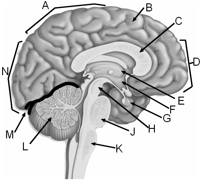
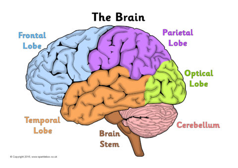
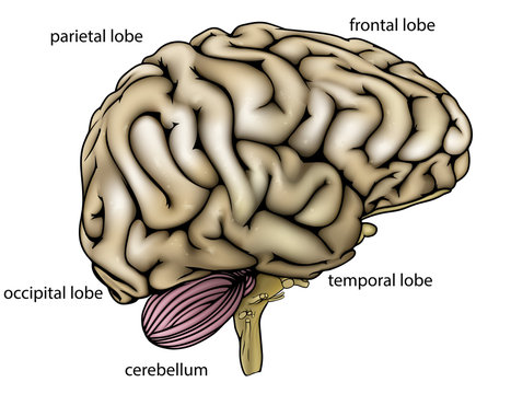
1,000+ Free The Brain & Brain Images - Pixabay 1,686 Free images of The Brain Related Images:brainmindpsychologythinkanatomyideasciencethoughtheadhuman Free the brain images to use in your next project. Browse amazing images uploaded by the Pixabay community. 1544259 brainmindpsychology 632119 artificial intelligence 656171 artificial intelligence 669161 questionquestionsman 1121258 Labeled Brain Model Diagram | Science Trends The cerebrum is the largest and most complex portion of the human brain. The cerebrum's function is to control our actions and thoughts, either conscious or unconscious, and responses to stimuli. The cerebrum itself is typically divided into four different lobes: the temporal lobe, the parietal lobe, the occipital lobe, and the frontal lobe. Brain: Anatomy, Pictures, Functions, and Conditions - Verywell Mind The brainstem is an area located at the base of the brain that contains structures vital for involuntary functions such as the heartbeat and breathing. The brain stem is comprised of the midbrain, pons, and medulla. 3 Midbrain The midbrain is often considered the smallest region of the brain. Human Brain Diagram Photos and Premium High Res Pictures - Getty Images 1,038 Human Brain Diagram Premium High Res Photos Browse 1,038 human brain diagram stock photos and images available, or start a new search to explore more stock photos and images. of 18 NEXT
Brain Labels Stock Illustrations – 243 Brain Labels Stock ...
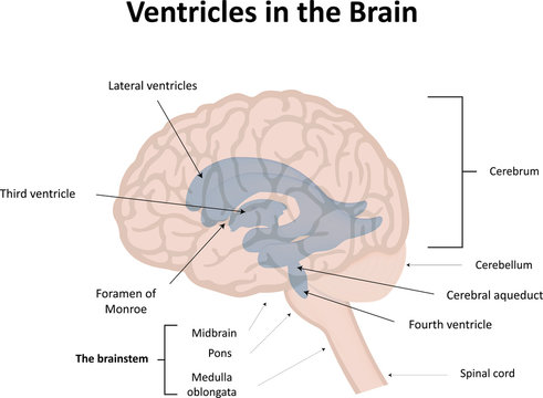
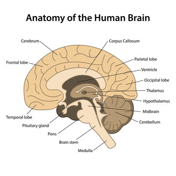
Diagram Of Brain with their Labelings and Detailed Explanation - BYJUS A well-labelled diagram of a human brain is given below for further reference. Structure And Function Of The Human Brain Parts Of The Human Brain The human brain is divided into three main parts: Forebrain. Midbrain. Hindbrain. These three main parts comprises many small parts. Forebrain The forebrain is also called as Prosencephalon.
Label the brain - Teaching resources
Low-functioning autism - Wikipedia Low-functioning autism (LFA) is a degree of autism marked by difficulties with social communication and interaction, challenging behavior, and differences in social or emotional reciprocity.Sleep problems, aggression, stereotypical, and self-injurious behavior are also common symptoms. LFA is not a recognized diagnosis in the DSM-5 or ICD-10, as neither …

Lab 10: Brain Labeling Diagram | Quizlet
2,000+ of the Best Brain Pictures for Free [HD] - Pixabay 2,000 Pictures of Brain in HD Related Images: people human nervous system mind Pick the perfect brain picture for your project. HD to 4K quality, available for free on all devices!
Brain Label
Book Bin Labels Teaching Resources | Teachers Pay Teachers Browse book bin labels resources on Teachers Pay Teachers, ... Make your classroom decor both stylish and functional with real pictures! This set includes simple labels perfect for any decor theme!I have included themed labels as well as resource bin labels for you! ... The Primary Brain. 4.9 (280) $3.50. PPTX;
Brain Anatomy - Label the Brain Diagram - Human Body Science Clip Art / Clipart
Toward Sharing Brain Images: Differentially Private TOF-MRA Images With ... Additionally, we compared the images using the FID as proposed in previous studies (Haarburger et al., 2019; Coyner et al., 2022). The generated image-label pairs were evaluated by a U-Net for brain vessel segmentation adapted from Livne et al. . After training the GANs, 41,000 image-label pairs were generated.
Frontiers | 101 Labeled Brain Images and a Consistent Human ...
Browse Labels & Organizations - VGMdb VGMdb provides media, tracklist and artist information for video game soundtracks and anime music.

File:Brain diagram without text.svg - Wikimedia Commons
Selegiline Oral: Uses, Side Effects, Interactions, Pictures ... - WebMD Find patient medical information for selegiline oral on WebMD including its uses, side effects and safety, interactions, pictures, warnings and user ratings.

AP 1 Lab 12- Brain Labeling Diagram | Quizlet
Free Classroom Labels Teaching Resources | Teachers Pay … This free sample pack includes four polka dot themed labels for classroom notebooks. Use them to clearly label your students’ notebooks in language arts, math, social studies, and science! Each label is 4 inches by 3.3 inches. Just print the labels on any brand of sticky labels that come in this size (six labels per sheet), peel, and stick!

Exterior Brain labeling Quiz worksheet
Brain: Atlas of human anatomy with MRI - e-Anatomy - IMAIOS Anatomy of the brain (MRI) - cross-sectional atlas of human anatomy. The module on the anatomy of the brain based on MRI with axial slices was redesigned, having received multiple requests from users for coronal and sagittal slices. The elaboration of this new module, its labeling of more than 524 structures on 379 MRI images in three different ...

Label Each Part of the Brain | MS in African Americans ...
Parts of the brain: Learn with diagrams and quizzes | Kenhub Labeled brain diagram First up, have a look at the labeled brain structures on the image below. Try to memorize the name and location of each structure, then proceed to test yourself with the blank brain diagram provided below. Labeled diagram showing the main parts of the brain Blank brain diagram (free download!)
2,823 Labeled brain anatomy Images, Stock Photos & Vectors ...
Haloperidol Oral: Uses, Side Effects, Interactions, Pictures Find patient medical information for haloperidol oral on WebMD including its uses, side effects and safety, interactions, pictures, warnings and user ratings.
Solved] Label the parts of the human brain and describe their ...
Parts of the Brain: Structures, Anatomy and Functions The brain is a 3-pound organ that contains more than 100 billion neurons and many specialized areas. There are 3 main parts of the brain include the cerebrum, cerebellum, and brain stem.The Cerebrum can also be divided into 4 lobes: frontal lobes, parietal lobes, temporal lobes, and occipital lobes.The brain stem consists of three major parts: Midbrain, Pons, and Medulla oblongata.
Basic Brain Diagram - resource - Imageshare
Youthful Brain Reviews: SCAM Ingredients? Don’t Buy Until Read … Mar 22, 2021 · As a result, the Youthful Brain formula came into existence to address and slow down brain aging and memory loss. According to the creator, the product might improve memory and regains brain focus .

Human Brain Diagram - Labeled, Unlabled, and Blank | Human ...
Amazon.com: Bulletproof Brain Octane C8 MCT Oil Travel Size ... Contains one 3-ounce bottle of premium Bulletproof Brain Octane C8 MCT Oil ; Pure MCT oil provides rapid mental and physical energy, supports cognitive function and helps control occasional cravings ; Brain Octane C8 MCT oil is extracted from 100% coconut oil, not palm oil, and rapidly converts into brain-powering, fat-burning ketone energy

Brain Crosssection With Labels Stock Illustration - Download ...
PDF Automatic labeling of MR brain images through extensible learning and ... Key words: atlas selection, brain MR images, image segmentation, learning, random forest 1. INTRODUCTION Accurate brain anatomy labeling is a crucial prerequisite for numerous clinical and research applications. However, man-ual labeling is a time-consuming task because labeling a set of MR brain image requires a specialist to work for 2 or
Labeled Brain Images – Browse 728 Stock Photos, Vectors, and ...
Human Brain Anatomy - Components of Human Brain with Images These are called the functions of brain. It is this part of the human body that serves as a control center for every activity in the body. In short, it is the brain that embodies the essence of mind and soul and death of this master organ literally means the death of entire body and the end of life. Brain: The Center of CNS:

Colored And Labeled Human Brain Diagram Stock Illustration ...
Picture of the Brain - WebMD Brain Tests. Computed tomography (): A scanner takes multiple X-rays, which a computer converts into detailed images of the brain and skull.Magnetic resonance imaging (): Using radio waves in a ...
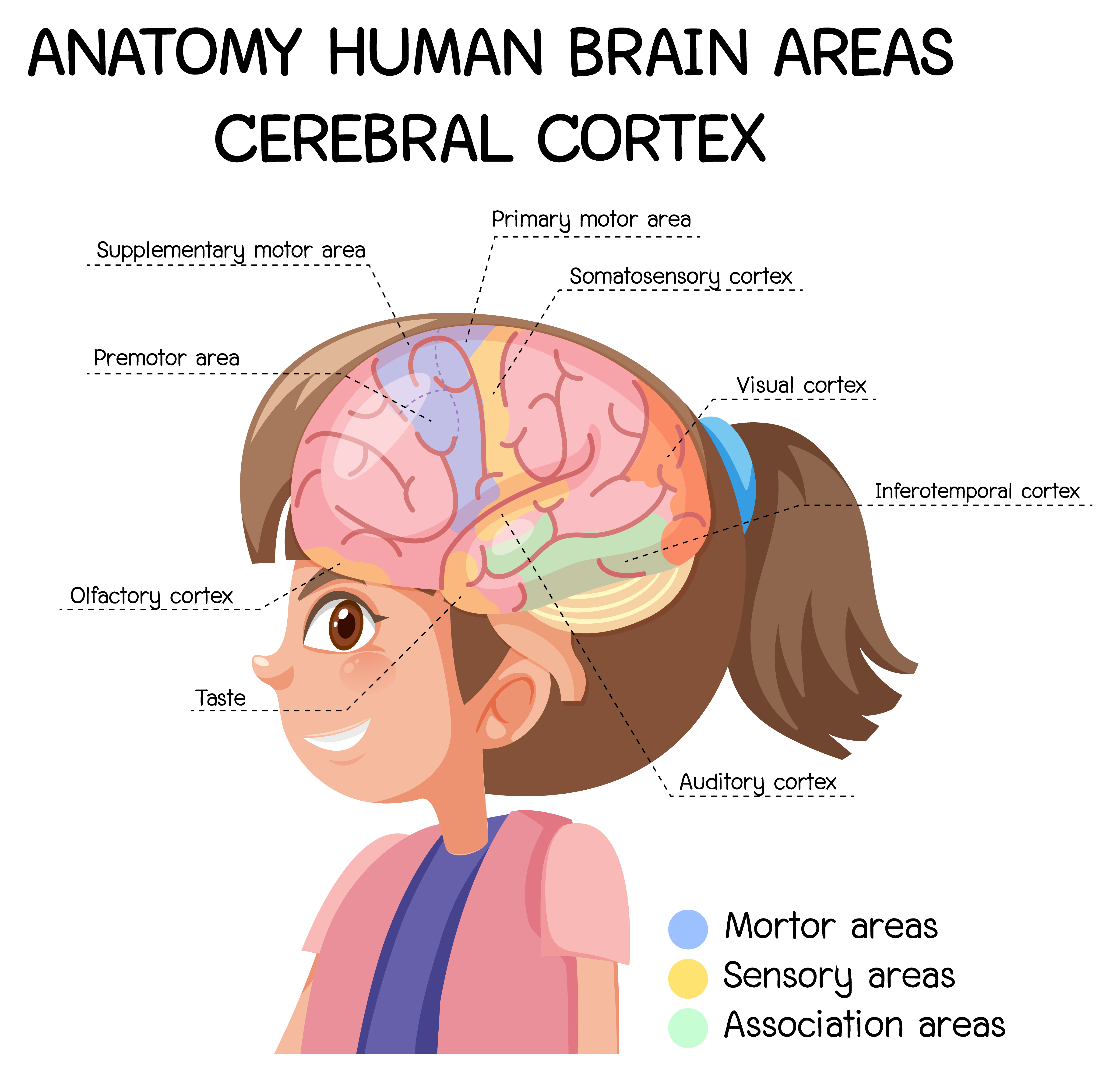
Anatomy human brain areas cerebral cortex with label 1846632 ...
101 labeled brain images and a consistent human cortical ... - PubMed 101 labeled brain images and a consistent human cortical labeling protocol Abstract We introduce the Mindboggle-101 dataset, the largest and most complete set of free, publicly accessible, manually labeled human brain images.
Label the Parts of the Brain
3D Brain Image Gallery - BrainHQ from Posit Science 3D Brain Image Gallery A group of Harvard scientists has developed new methods of magnetic resonance imaging (MRI) to look more closely inside the human brain. The resulting images are stunning!
Label the Brain Worksheets (SB11585) - SparkleBox
How to Create Labels in Microsoft Word (with Pictures) - wikiHow Jan 18, 2020 · Obtain the labels you need. Labels come in different sizes and for different purposes, for everything from regular, no. 10 envelopes to legal-sized mailings and CD covers. Get the labels that are best-suited for your project.

Midsaggital Brain w/ labels Diagram | Quizlet
Brain Label | Human anatomy and physiology, Ap biology, Basic anatomy ... Brain Label. Image of the brain showing its major features for students to practice labeling. Answers are included. Biologycorner. 18k followers ... Find Structure Human Brain Section Schematic Vector stock images in HD and millions of other royalty-free stock photos, illustrations and vectors in the Shutterstock collection. Thousands of new ...
Human Brain with Labels stock illustration. Illustration of ...
Labeling Brain Structures - John Muschelli 1 Labels in template space. In Processing Within-Visit MRI, we registered the T1 image to the Eve template using a non-linear registration (SyN) (Avants et al. 2008). Also, we applied this transformation to the intensity-normalized T1, T2, and FLAIR images, so that these image are located in the same space as the Eve atlases. We can overlay the ...

29,181 Brain labelled Images, Stock Photos & Vectors ...
PDF Automatic labeling of MR brain images by hierarchical learning of atlas ... Accurate brain anatomy labeling is a task of pivotal importance to region-based analysis of MR brain images. It can be further applied to the research and clinical studies, such as for facilitating diagnosis, guiding treatment, and monitoring disease progression.1 Since it is labor-intensive and impractical to label a large set of 3D MR images
Labeled Brain Images – Browse 728 Stock Photos, Vectors, and ...
Parts of the Brain Activity for Kids, Brain Diagram, and Worksheets for ... PARIETAL LOBES - The parietal lobe provides sensory information to the brain including touch, pain and temperature. OCCIPITAL LOBES - The occipital lobe processes and interprets everything we see TEMPORAL LOBES - The temporal lobe controls emotions and short-term memory
Brain Clipart With Labels, HD Png Download , Transparent Png ...
101 Labeled Brain Images and a Consistent Human Cortical Labeling ... We selected 101 T 1-weighted brain MR images that are: (1) publicly accessible with a non-restrictive license, (2) from healthy participants, (3) of high quality to ensure good surface reconstruction, and (4) part of a multi-modal acquisition ( T 2*-weighted, diffusion-weighted scans, etc.).

Human brain with labels, illustration Stock Photo - Alamy
Sheep Brain Label
External Brain Anatomy – Foundations of Neuroscience

How to draw human brain/draw and label brain diagram/draw labelled diagram of brain/brain diagram

Labeling the brain (Exam 1) Flashcards | Quizlet
2,823 Labeled brain anatomy Images, Stock Photos & Vectors ...
Human Brain Anatomy Sagittal Section Labels Stock ...

How to draw human brain/ draw labelled diagram of brain/brain diagram/draw and label brain diagram

Brain Labeling Diagram | Quizlet

Brain Labeling | The brain for kids, Writing a persuasive ...
Label the Brain Worksheets (SB11585) - SparkleBox
Labeled Brain Images – Browse 728 Stock Photos, Vectors, and ...
How The Brain Works! - Lessons - Blendspace
2,823 Labeled brain anatomy Images, Stock Photos & Vectors ...
Parts of the brain without labels" Poster for Sale by Kru22 ...
Post a Comment for "40 brain pictures and labels"